Vision Therapy for Concussion & Traumatic Brain Injury: Recovery Guide (2026)
This article is educational and is not a diagnosis or treatment plan. Clinical claims should be interpreted with a qualified eye-care professional. See our evidence library and clinical standards for methodology and source context.
Concussion & the Visual System: Why Vision Is Affected
Concussion is a mild traumatic brain injury (mTBI) that disrupts the brain’s normal functioning. Because more than 60% of the brain’s neural pathways are involved in vision, concussion frequently causes significant visual disturbances — even when the eyes themselves are structurally normal. Structured rehabilitation for these deficits is the focus of neuro-optometric vision therapy.
The visual system is uniquely vulnerable to concussion for several reasons. Visual processing requires the coordinated function of cranial nerves (II, III, IV, VI), the cerebellum, brainstem, and widespread cortical networks. The shearing forces that occur during a concussion can damage axons throughout these pathways, disrupting the timing and coordination required for clear, single, comfortable vision. Common mechanisms of injury include sports collisions, motor vehicle accidents, falls, and blast injuries among military personnel.
Research published in the Journal of Neuro-Ophthalmology (2024) found that up to 70% of concussed patients report at least one visual symptom, and approximately 40% meet the diagnostic criteria for a binocular vision disorder that persists beyond the acute injury phase. Despite this high prevalence, post-concussion vision problems are frequently overlooked or misattributed to other causes, delaying appropriate treatment.
Vision Problems After Concussion & TBI
Post-concussion vision problems manifest across several domains. The most common complaints include:
Binocular vision disorders — Convergence insufficiency (CI) is the single most common vision disorder following concussion, affecting an estimated 40–50% of patients with persistent symptoms. The eyes struggle to maintain alignment during near tasks, causing double vision (diplopia), eye strain, and difficulty reading. Divergence excess and basic exophoria are also frequently observed. Our convergence insufficiency guide provides a detailed overview of CI diagnosis and management.
Accommodative dysfunction — The ability to focus clearly at different distances (accommodation) is commonly impaired after concussion. Patients report fluctuating blur, difficulty shifting focus from distance to near, and visual fatigue that sets in rapidly during sustained near work.
Oculomotor dysfunction — The smooth tracking (pursuits) and rapid eye movements (saccades) that are essential for reading and scanning the environment are often disrupted. Patients may lose their place while reading, have difficulty tracking moving objects, and experience nausea or dizziness in visually complex environments such as grocery stores.
Photosensitivity — Heightened sensitivity to light is a hallmark of post-concussion syndrome. Bright environments, fluorescent lighting, and screen glare can trigger headache, eye pain, and symptom exacerbation.
Visual-vestibular mismatch — The integration between vision and the vestibular (balance) system is frequently disrupted. This creates symptoms of dizziness, unsteadiness, and motion sensitivity, especially in visually busy environments or when moving through space.
Visual processing speed deficits — Many patients report that visual information feels slower or more effortful to process after their injury. Scanning a page, interpreting a chart, or navigating a new environment requires more mental effort than before the concussion.
Why Standard Eye Exams Miss Post-Concussion Vision Issues
One of the most frustrating experiences for concussion patients is being told their eyes are "fine" by a standard eye exam, despite ongoing visual symptoms. This happens because standard eye exams assess only visual acuity and eye health — not the functional aspects of vision that concussion affects.
A standard eye chart test measures the ability to see letters at a distance (Snellen acuity). It does not assess:
- How well the eyes work together as a team (binocular vision)
- The ability to maintain convergence during sustained near tasks
- Accommodative flexibility (focus shifting speed and accuracy)
- Saccadic and pursuit eye movement accuracy
- Visual processing speed under cognitive load
- Visual-vestibular integration
A comprehensive neuro-optometric evaluation is required to identify these deficits. This is distinct from both a routine eye exam and a neurological examination — it sits at the intersection of the two disciplines. The same diagnostic gap exists for other conditions like amblyopia and functional vision disorders, where standard screening tools are insufficient.
The Neuro-Optometric Evaluation for Post-Concussion Vision
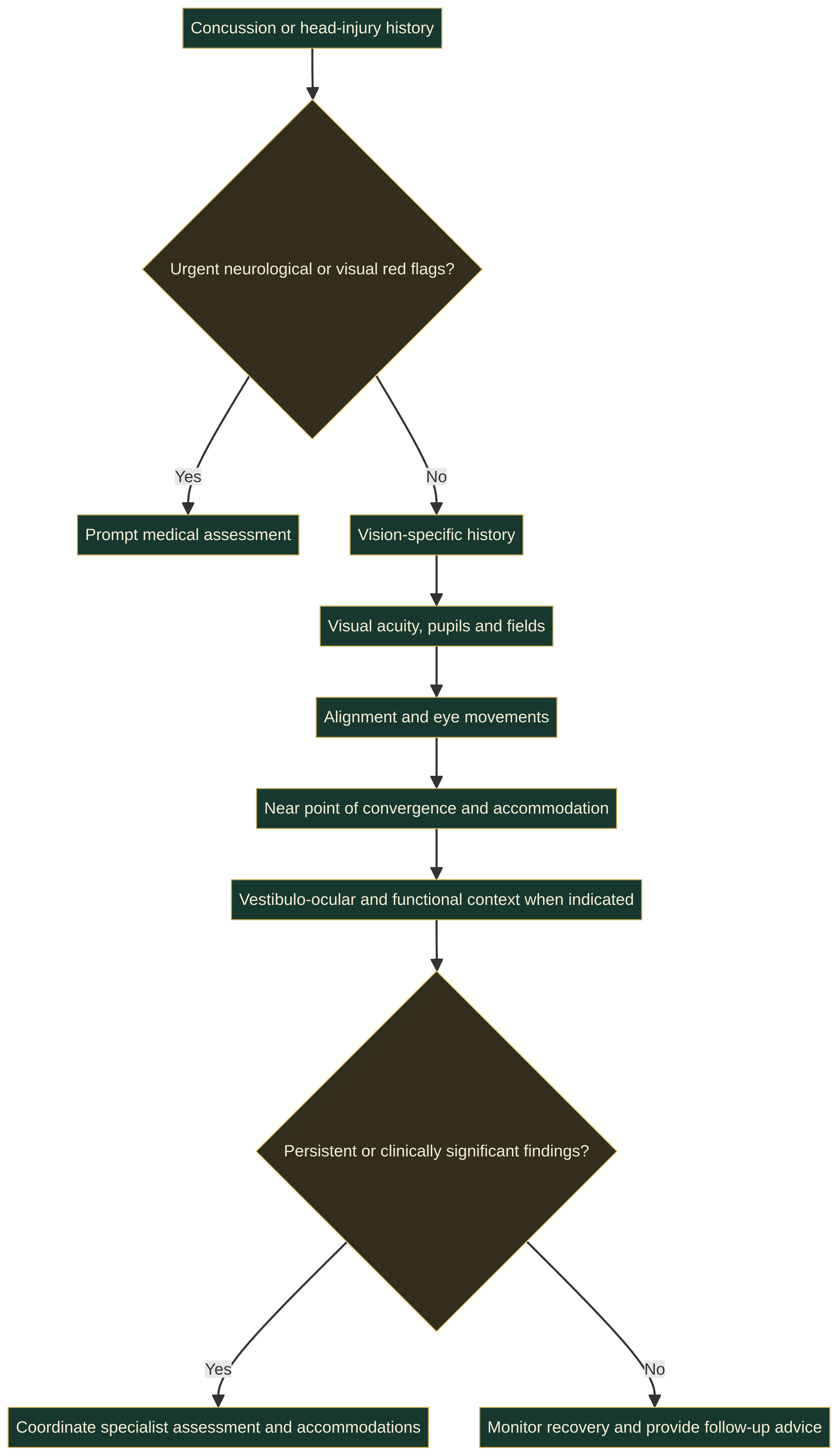
A neuro-optometric evaluation for concussion patients includes several components not found in a standard eye exam:
Comprehensive case history — The doctor will ask about the mechanism of injury, symptom onset, aggravating factors (reading, screens, driving, crowded environments), and prior treatments. Standardised symptom inventories such as the Visio-Vestibular Symptom Questionnaire (VVSQ) or the Post-Concussion Symptom Scale (PCSS) help quantify impairment.
Binocular vision assessment — Near point of convergence (NPC), positive and negative fusional vergence ranges, and cover testing at distance and near. A receded NPC (>10 cm) is a hallmark finding and a strong predictor of persistent post-concussion symptoms.
Accommodative testing — Accommodative amplitude, facility (plus/minus lens flipper testing), and lag of accommodation are measured to identify accommodative insufficiency or infacility.
Oculomotor testing — The King-Devick (K-D) test, NSUCO oculomotor test, and Developmental Eye Movement (DEM) test quantify saccadic accuracy, speed, and the presence of head movements during tracking. The K-D test is also increasingly used as a sideline concussion screening tool.
Visual-vestibular integration — Dynamic visual acuity (DVA) testing, gaze stability during head movement, and motion sensitivity testing assess the integrity of the vestibulo-ocular reflex (VOR).
Photosensitivity assessment — Tolerance to light intensity and flicker is evaluated using standardised light exposure protocols.
Vision Therapy for Post-Concussion Syndrome
Vision therapy is the primary evidence-based treatment for post-concussion visual dysfunction. Unlike rest alone, which leaves neural pathways to heal suboptimally, vision therapy actively retrains the brain to recover accurate visual processing through structured, progressive exercises.
The treatment approach differs from standard vision therapy in important ways. Post-concussion patients often have reduced exercise tolerance and must start with shorter sessions (10–15 minutes) at lower intensity, gradually progressing as symptoms improve. This sub-symptom threshold approach is adapted from vestibular therapy protocols and is essential for avoiding symptom exacerbation.
A typical post-concussion vision therapy programme runs 12–24 weeks with the following phases:
Phase I: Foundation (Weeks 1–4) — Focus on monocular oculomotor control, basic accommodative facility, and vestibulo-ocular reflex (VOR) exercises. The goal is to build a stable foundation without provoking symptoms.
Phase II: Binocular Integration (Weeks 5–12) — Convergence and divergence training, vergence facility, and binocular accommodative facility are introduced. This phase directly addresses convergence insufficiency, the most common post-concussion binocular disorder.
Phase III: Functional Application (Weeks 13–20) — Visual processing speed training, visual-vestibular integration under dynamic conditions, and visual-motor integration tasks prepare the patient for return to school, work, and sport.
Phase IV: Maintenance (Weeks 20–24) — Independent home exercise programme with periodic re-assessment to ensure gains are maintained and to address any residual deficits.
Key VT Modules for Concussion & TBI Recovery
GO VISION THERAPY provides specialised modules targeting the visual deficits most commonly seen after concussion:
Jump Ductions — Rapid vergence changes between distance and near targets to improve convergence amplitude and facility. Critical for treating post-concussion convergence insufficiency.
Variable Vergence — Graded vergence demand allows clinicians to precisely control the convergence requirement and progressively load the vergence system as the patient improves. This is especially important for sub-symptom threshold rehabilitation.
Anti-Saccade Training — Patients must inhibit a reflexive glance toward a peripheral target and instead look in the opposite direction. This executive function task is particularly sensitive to frontal lobe injury and is a core component of cognitive vision rehabilitation.
Visual Flow Stimulation — Optokinetic and full-field motion stimulation helps recalibrate the visual-vestibular mismatch that causes motion sensitivity and dizziness in visually complex environments.
3D Multiple Object Tracking (3D MOT) — Tracking multiple moving objects in three-dimensional space exercises attention, visual processing speed, and divided visual field awareness — all of which are commonly affected after concussion.
Tachistoscope — Brief visual presentations at controlled exposure durations build visual processing speed and accuracy, which are frequently reduced after brain injury. Our Digital Toolbox includes full details on each module.
Research & Evidence for VT in Concussion Recovery
The evidence base for vision therapy in post-concussion rehabilitation has grown substantially in recent years:
A 2023 systematic review in Optometry and Vision Science (Gallaway et al.) analysed 14 studies on vision therapy for concussion-related binocular vision disorders. The review found strong evidence that office-based vergence and accommodative therapy significantly improves near point of convergence, positive fusional vergence, and accommodative amplitude in post-concussion patients, with 73–88% achieving clinically significant improvement.
A landmark randomised controlled trial by Scheiman et al. (2021) published in Optometry and Vision Science compared office-based vision therapy to home-based pencil push-ups for post-concussion convergence insufficiency. The office-based therapy group showed significantly better outcomes on NPC, PFV, and symptom scores (CISS) compared to the home-based group, with effects sustained at 6-month follow-up. This mirrors findings in the CITT trials for convergence insufficiency in the general population.
A 2022 retrospective study in the Journal of Neuro-Optometry examined 98 paediatric patients with sports-related concussion who received vision therapy. Results showed that 78% had a complete resolution of visual symptoms within 12 weeks, and the average time to return to school full-time was reduced from 28 days (historical controls) to 16 days in the VT-treated group.
The American Optometric Association (AOA) and the Neuro-Optometric Rehabilitation Association (NORA) both endorse vision therapy as a cornerstone of multidisciplinary concussion rehabilitation. The AOA’s 2025 Clinical Practice Guideline for concussion management recommends a comprehensive eye examination including binocular vision assessment for all patients following concussion, with vision therapy as the first-line treatment for diagnosed visual dysfunction.
GO VISION THERAPY for Concussion & TBI Recovery
GO VISION THERAPY offers a purpose-built platform for post-concussion vision rehabilitation. Our digital approach addresses several unique needs of concussion patients:
Precise stimulus control — Every exercise parameter (vergence demand, exposure duration, contrast, speed) is computer-controlled with millisecond precision. This allows clinicians to start at sub-symptom threshold levels and incrementally progress with confidence.
Remote monitoring — Patients complete therapy sessions at home while clinicians monitor compliance, performance, and symptom reports in real time. This is particularly beneficial for concussion patients who may have reduced tolerance for travel and clinic visits.
Automated progress tracking — The platform generates detailed reports on NPC change, vergence ranges, processing speed metrics, and symptom trends over time. These objective outcome measures support return-to-play, return-to-learn, and return-to-work decisions.
Integrated symptom monitoring — Patients log pre- and post-session symptom scores, allowing clinicians to identify activity thresholds and adjust programmes accordingly. This feature is critical for the sub-symptom threshold approach that defines best-practice concussion rehabilitation.
For optometrists and neuro-optometric rehabilitation specialists, GO VISION THERAPY extends clinical capacity beyond the office, enabling effective home-based therapy with the same precision and control as in-clinic exercises. Explore our software platform for full details.
When to Seek a Neuro-Optometric Evaluation
If you or someone you care for has experienced a concussion or TBI and experiences any of the following, a comprehensive neuro-optometric evaluation is strongly recommended:
- Persistent headaches during or after reading, screen use, or visually demanding tasks
- Double vision or blurred vision that comes and goes
- Difficulty reading for more than a few minutes without discomfort
- Feeling overwhelmed, dizzy, or anxious in visually busy environments (supermarkets, traffic, crowded rooms)
- Sensitivity to light that did not exist before the injury
- Poor balance or increased motion sickness when walking or riding in vehicles
- Difficulty concentrating on visual tasks that were previously easy
- Children struggling to return to school because of reading or classroom-related fatigue
Early intervention yields the best outcomes. While some visual symptoms improve spontaneously in the first 2–4 weeks post-injury, persistent symptoms lasting beyond 4 weeks are unlikely to resolve without targeted treatment. The window of neuroplasticity is optimal in the first 3–6 months, but vision therapy remains effective even in chronic post-concussion syndrome, including cases lasting years after injury.
Patients already receiving care for post-concussion syndrome from a neurologist, physiatrist, or vestibular therapist should ask about adding a vision therapy evaluation to their care team. Vision rehabilitation works best as part of a coordinated multidisciplinary approach. Our guide to vision therapy after neurological injury provides additional context on the role of VT in brain injury recovery.
Frequently Asked Questions
Acute concussion (first 1–2 weeks) is typically managed with relative rest. After this initial period, if visual symptoms persist, a neuro-optometric evaluation should be scheduled. Vision therapy can begin as early as 2–4 weeks post-injury for patients with stable symptoms. Early intervention within the first 3 months yields the best outcomes, but late intervention (even years post-injury) can still be effective.
Yes, with proper clinical supervision. Research shows that office-based vergence/accommodative therapy is more effective than unsupervised home exercises, but digital platforms like GO VISION THERAPY bridge this gap by providing office-quality precision in a home setting. The key is that therapy must be guided by a clinician who sets parameters, monitors progress, and adjusts the programme as the patient improves. Unsupervised pencil push-ups alone are proven inferior to structured, clinician-guided therapy.
Coverage varies, but vision therapy for post-concussion visual dysfunction is increasingly covered by medical insurance when diagnosed and prescribed by a qualified professional. The medical necessity is well-established, and GO VISION THERAPY provides documentation and outcome reports to support claims. Many patients also use health savings accounts (HSAs) or flexible spending accounts (FSAs) to cover treatment.
The typical treatment course for post-concussion vision therapy is 12–24 weeks, with patients attending weekly in-office or telehealth-guided sessions and performing home exercises 5–6 days per week for 15–30 minutes per session. Many patients begin to notice improvement within 4–6 weeks. The duration depends on injury severity, symptom tolerance, treatment consistency, and the specific visual deficits identified.
Yes. Visual-vestibular integration training is one of the core components of post-concussion vision therapy. Exercises that combine eye movements with head movements (gaze stability, VORx1/VORx2, optokinetic stimulation) retrain the brain to integrate visual and balance information correctly. This is often combined with vestibular therapy for optimal outcomes. Most patients report significant reduction in visually induced dizziness after 6–12 weeks of targeted therapy.
Absolutely. Children are particularly good candidates for vision therapy because of their high degree of neuroplasticity. Paediatric concussion patients who receive vision therapy return to school and sport significantly faster than those who do not. The children’s vision therapy guide provides more information on how VT is adapted for younger patients. Parents should watch for signs such as declining grades, avoidance of reading, or increased screen time complaints after any head injury.
Yes — in fact, most concussions do not involve loss of consciousness. The forces required to cause a concussion (rapid acceleration-deceleration of the head) are sufficient to stretch and damage axons in the visual pathways even when the person remains conscious throughout. Vision problems can develop after even a seemingly mild head impact, and their severity often correlates poorly with the apparent severity of the injury. Any blow to the head followed by visual symptoms warrants evaluation.
Post-concussion vision therapy follows the same principles as standard VT but with important modifications: (1) sub-symptom threshold exercise intensity to avoid symptom flare-ups, (2) heavier emphasis on visual-vestibular integration, (3) graded return-to-functional demands (school, work, sport), and (4) close coordination with other rehabilitation providers (vestibular therapists, occupational therapists, physiatrists). The exercises themselves — vergence, accommodation, oculomotor training — are shared with standard VT, but the dosing and progression are tailored to the post-concussion brain.
- Alvarez TL et al. CONCUSS randomised clinical trial of vergence/accommodative therapy for concussion-related symptomatic convergence insufficiency. British journal of sports medicine, 2026. PubMed (PMID 41033748)
- Chen AM et al. Scoping review: Intervention for ocular motor disorders in children and adults with mild traumatic brain injury. Optometry and vision science : official publication of the American Academy of Optometry, 2025. PubMed (PMID 39951336)
- Harmon KG et al. Validity of the King Devick Test for Acute Diagnosis of Concussion in NCAA Division I Athletes. Sports health, 2026. PubMed (PMID 40625038)
- Mavroudis I et al. Neuro-visual pathway after mild traumatic brain injury: a systematic scoping review of symptoms, objective testing, and rehabilitation. Acta neurologica Belgica, 2026. PubMed (PMID 42287492)
Vision Therapy for Concussion & TBI Recovery
GO VISION THERAPY provides clinicians and patients with a purpose-built platform for post-concussion vision rehabilitation. Monitor progress, adjust parameters remotely, and achieve measurable outcomes.